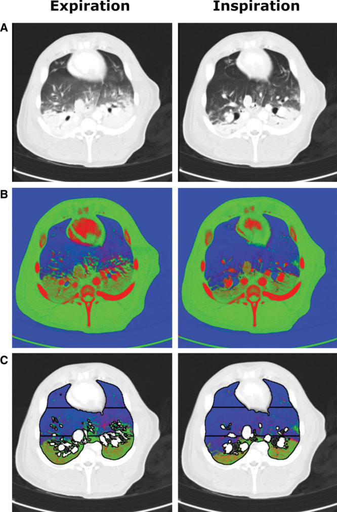
Figure 2.

Example source and post-processed images of a single juxtadiaphragmatic slice at positive end-expiratory pressure 5 cm H2O of pig’s thorax during iodine infusion using the dual-energy CT (DECT) algorithm. A, Composite source images representing a 30:70 merge of 80 kVp and 140 kVp images displayed using standard CT lung windows. B, Results of the DECT three-material differentiation algorithm for gas (blue), soft tissue (green), and iodinated blood (red) volume fractions. C, The DECT images following segmentation to include only lung parenchyma with the three gravitational regions of interest displayed. Typical expiration and inspiration images are shown in each case. A gravitational effect was seen within the slice with soft tissue and iodinated blood concentrated toward the dependent regions, with a reduction in volume fractions of these materials in inspiration.
