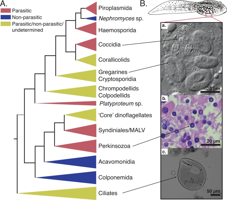
Fig 2. Schematic tree of the Alveolata superphylum illustrated with some examples of tadpole infectious agents.
A. Schematic diagram of the relationships among the three main lineages of the Alveolata superphylum based on rDNA phylogeny (not to scale), with parasitic and nonparasitic lineages indicated. Dotted line for the basal branch is hypothetical. Adapted from Mickhailov and colleagues (2014) [15]. B. Micrographs of tadpole liver and intestine samples infected by protists belonging to the Alveolata superphylum. a. Light microscopy of macrophages containing several oocysts of both Nematopsis temporariae (Gregarines) and Goussia noelleri (Coccidia) from tadpole liver samples of Rana dalmatina, fresh mounts, NIC [6] b. Histological section of infected liver tissue samples from a River frog (Rana heckscheri) tadpole mass mortality event in southwestern Georgia (USA) in 2006, stained with hematoxylin–eosin (Yabsley, unpublished). c. Light microscopy of putatively commensal ciliate Balantidium sp. from tadpole intestine samples of Bombina bombina, fresh mounts, NIC (Jirků, unpublished). The tadpole drawing is a free public domain vector cliparts (available on www.clker.com). NIC, Nomarski interference contrast.

