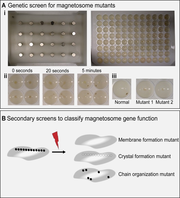
Fig 2. Magnetic screening technique.
(A) (i) 24-pin magnetic plate (left) and 96-well plate of AMB-1 cells (right) used in Komeili and colleagues. [34]. (ii) Movement of AMB-1 cells on magnetic plate at 0 seconds, 20 seconds, and 5 minutes. (iii) Phenotype of normal magnetic cells (left) and two representative, nonmagnetic mutants (right). (B) Diagram of secondary screens to classify magnetosome mutants.

