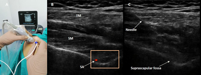
Figure 1.
A) The positioning of the linear ultrasound transducer and radiofrequency electrode. B) Scanning of the suprascapular nerve with linear ultrasound probe; trapezius muscle (TM), suprascapular muscle (SM), suprascapular notch, and color Doppler imaging of the suprascapular artery (SA). C) Real-time imaging of the needle insertion under ultrasonographic guidance.

