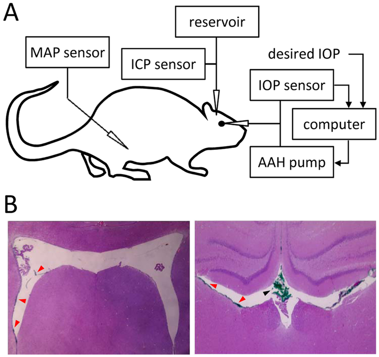
Figure 1.
Experimental setup. A, Intraocular pressure (IOP), intracranial pressure (ICP), and mean arterial pressure (MAP) were simultaneously recorded with separate pressure sensors via cannulas inserted in the anterior chamber of the eye, cerebral ventricles, and femoral artery, respectively. The IOP cannula was also connected to a pump that infused artificial aqueous humor (AAH) under computer control in order to measure conventional outflow facility. The ICP cannula was also connected to a variable-height reservoir of physiological saline in order to manipulate ICP level. B, Coronal tissue sections of a rat brain perfused with green tracer dye through the cannula. Sections are ~1 mm apart in the rostrocaudal direction. Red and black arrowheads indicate clumps of dye molecules in the lateral ventricles and dorsal third ventricle, respectively. No tracer was found in brain tissue in these or other sections.

