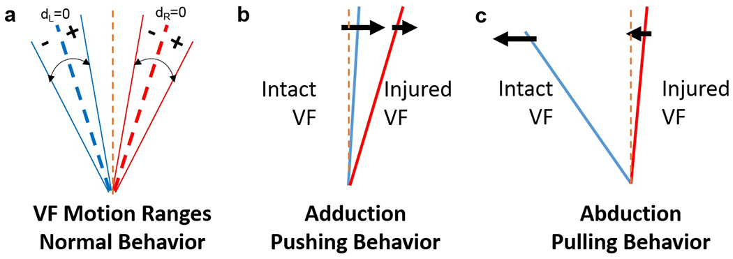
Figure 4.

Analysis of VF motion behavior. (a) Left and right VF motion ranges; center dashed blue and red lines represents motion midline for each VF (i.e., dL= displacement of left VF = 0; dR= displacement of right VF = 0). (b) VF motion during pushing: The intact VF contacts the injured VF to push it laterally in the same direction during VF adduction. (c) VF motion during pulling: The intact VF pulls the injured VF medially in the same direction during abduction. Left VF (blue line) represents the healthy, intact VF; right VF (red line) represents the injured VF. Orange dashed line = glottal midline.
