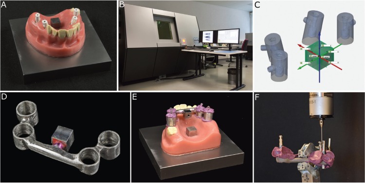
Figure 1.
A: Implant master model (IMM) with tightened scanbodies on the premolar (15° inclination) and molar (0° inclination) implants and the reference cube in the middle of the palate. B: Multisensor coordinate measuring machine using X-ray tomography (TomoScope S). C: Reference file (patient equivalent) with the implant-abutment interface centers (IAICs) from the computed tomography (green points). D: Custom made measuring aid (CMA) with the inherent reference cube. E: CMA coded on the IMM. F: Measurement of the transferred CMA with tightened laboratory analogues using the coordinate measuring machine (CMM).

