. 2020 Feb 13;9:e51992. doi: 10.7554/eLife.51992

Figure 1. Recombinant human tubulin lacking GTPase activity strongly nucleates microtubules.

(a) The evolutionarily conserved catalytic glutamic acid of α-tubulin (E, pink). (b) Coomassie Blue-stained SDS gel of purified wildtype (WT) and E254A mutant recombinant human tubulin. (c) HPLC chromatograms of pure GDP and GTP compared to nucleotides extracted from microtubules polymerized from porcine brain tubulin or wildtype and E254A mutant human tubulin. (d) Quantification of the nucleotide content of different microtubules. Bar graphs depict the means of 3 independent experiments, error bars represent standard deviation (SD). (e) iSCAT microscopy images of unlabeled wildtype human tubulin (12 µM) growing from immobilized GMPCPP-stabilized biotinylated seeds (GMPCPP-seeds). (f) Kymographs showing wildtype microtubule growth, conditions as in (e). (g, h) iSCAT microscopy images showing (g) lack of microtubule nucleation at 5 µM wildtype tubulin and (h) strong nucleation at 4 µM E254A tubulin at a surface with an immobilized rigor kinesin (Kin1rigor). Bottom: magnified E254A microtubule views. Scale bars as indicated, time is always min:sec.

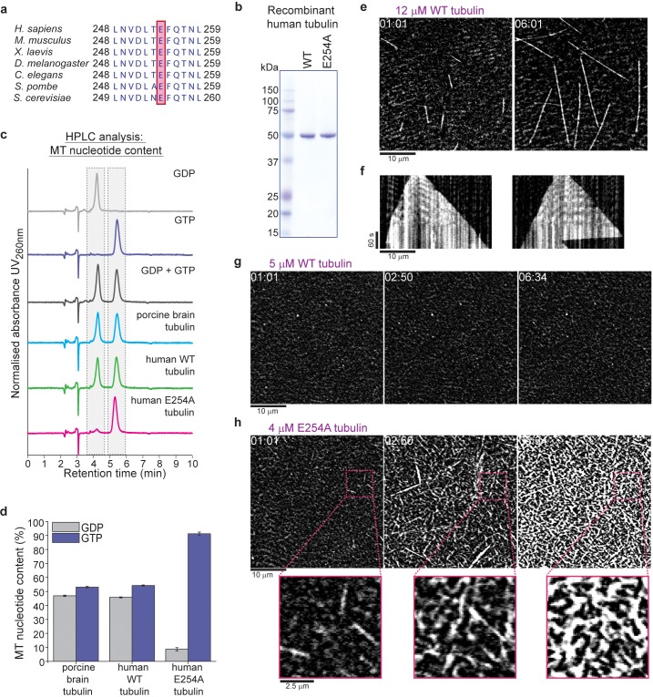
Figure 1.

Figure 1—figure supplement 1. Expression and purification of recombinant human α/β tubulin.

Figure 1—figure supplement 1.

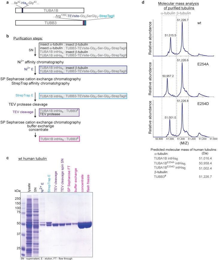
(a) Schematic of the human TUBA1B α-tubulin and TUBB3 β-tubulin expression constructs. (b) Flow chart of the purification steps to obtain recombinant human α/β-tubulin from insect cells. The hash (#) sign on TUBB3 marks the TEV-protease cleavage site. (c) Coomassie Blue-stained SDS gel showing the step-wise purification of recombinant wildtype (wt) human tubulin. Coloring of the labels as for the corresponding purification steps in the flow chart (b). (d) Molecular mass determination of purified recombinant tubulin subunits confirms the lack of insect cell tubulin contamination and uncleaved TUBB3. Note that the predicted masses for TUBA1B and its mutants correspond to N-terminally acetylated versions of the protein. This is a common modification of proteins expressed in insect cells.

Figure 1—figure supplement 2. iSCAT microscopy of unlabeled porcine brain microtubules.

Figure 1—figure supplement 2.

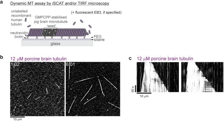
(a) Schematic of a dynamic microtubule assay. (b) iSCAT microscopy images of unlabeled porcine brain microtubules growing from GMPCPP-seeds at 12 µM porcine brain tubulin. (c) Kymographs showing the characteristic fast plus and slow minus end growth of dynamic porcine brain microtubules. Conditions as in (b). Scale bars as indicated, time is in min:sec.