. 2020 Jan 30;9:e53243. doi: 10.7554/eLife.53243

Figure 3. The structures of uninhibited IMPDH2 filaments.

(A) Cryo-EM of IMPDH2 filaments with both substrates (representative 2D class average). (B) We resolved two types of structures from IMPDH filaments: the consensus filament assembly interface, and various conformations of filament segments. (C) Cryo-EM density for the ATP/IMP/NAD+ consensus filament assembly interface, consisting of two tetramers bound back-to-back (dark and light green) (0.5 mM ATP, 2 mM IMP, 2 mM NAD+). (D) The filament assembly interface is mediated by the vertebrate-specific N-terminus, in particular a key bridge between Y12 and R356. (E) Cryo-EM density for the ATP/IMP/NAD+ fully extended filament segment. Opposing catalytic tetramers (dark and light green), are held separate by their symmetrically extended Bateman domains (dark and light pink). ATP (bright green) is resolvable in the Bateman domains. (F) In the fully extended Bateman domains, sites 1 and 2 are occupied by ATP, and site 3 is unformed. (G) Cryo-EM density for the best resolved ATP/IMP/NAD+ bent filament segment, in which the two catalytic tetramers are not parallel. (H) Filament segment bending results from asymmetric compression of Bateman domains. In this reconstruction, one protomer from each of the two tetramers is compressed, and allosteric site 3 is formed, but unoccupied (black asterisk). (I) Summary of the ATP/NAD+ cryo-EM dataset (2 mM ATP, 2 mM NAD+). The filament assembly interface is unchanged, and filament segments varied from fully extended, to bent, to fully compressed. In the absence of IMP, the flexible active site loops are disordered. (J) Summary of the ATP/IMP cryo-EM dataset (2 mM ATP, 3 mM IMP).

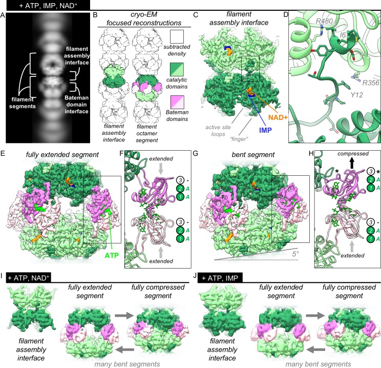
Figure 3—source data 1. Statistics of cryo-EM data collection, reconstruction and model refinement for the ATP/IMP/NAD+ dataset.
Figure 3—source data 2. Statistics of cryo-EM data collection, reconstruction and model refinement for the ATP/NAD+ dataset.
Figure 3—source data 3. Statistics of cryo-EM data collection, reconstruction and model refinement for the ATP/IMP dataset.

Figure 3.

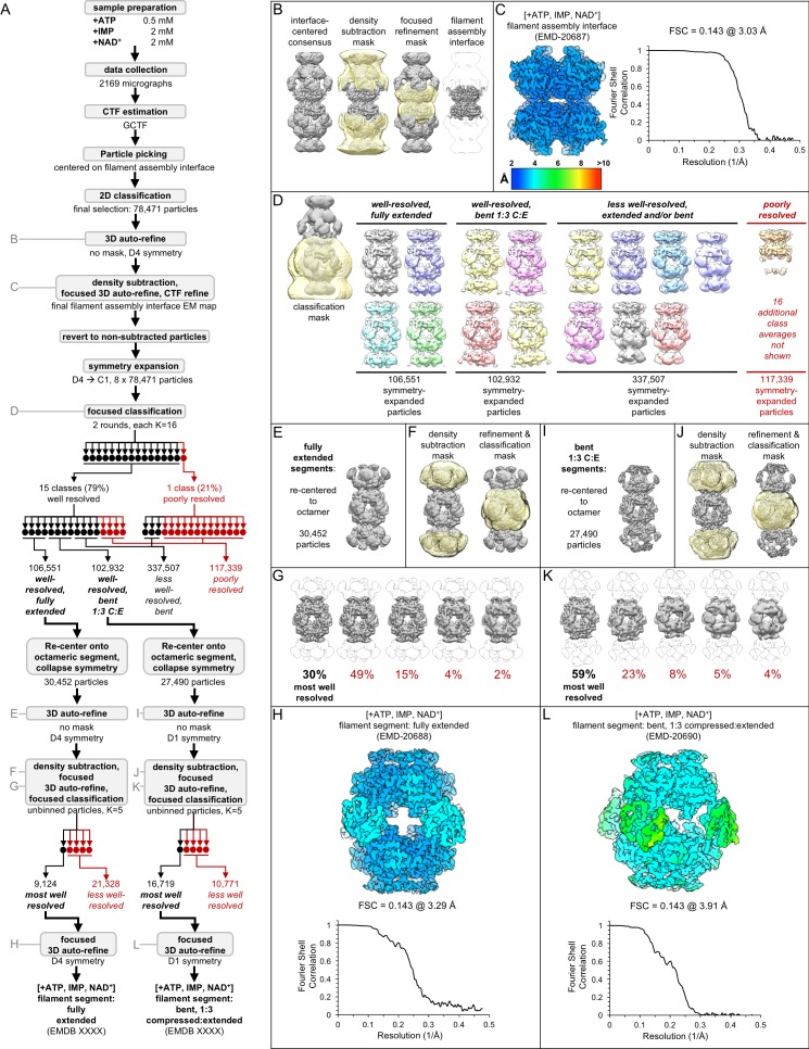
Figure 3—figure supplement 1. Image processing of the IMPDH2 +ATP, IMP, NAD+ cryo-EM dataset.

Figure 3—figure supplement 1.

Nucleotide concentrations for this dataset: 0.5 mM ATP, 2 mM IMP, 2 mM NAD+. (A) Flow chart summarizing data processing strategy. (B) Density subtraction and focused refinement of the consensus filament assembly interface. (C) Local resolution estimation and FSC curve (via relion postprocessing) for the ATP/IMP/NAD+ consensus filament assembly interface. (D) Final class averages from symmetry expanded classification of filament segments. (E) Unmasked refinement from all fully extended segments, pooled and re-centered. (F) Masks used for continued processing of fully extended segments. (G) Final classification of the best-resolved fully extended filament segment class H) Local resolution estimation and FSC curve for the ATP/IMP/NAD+ fully extended filament segment I-L) Same as E-H, but for the best-resolved ATP/IMP/NAD+ bent filament segment.
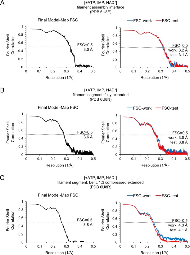
Figure 3—figure supplement 2. Model/Map FSC curves for the IMPDH2 +ATP, IMP, NAD+ cryo-EM dataset.

Figure 3—figure supplement 2.

Nucleotide concentrations for this dataset: 0.5 mM ATP, 2 mM IMP, 2 mM NAD+. For each structure, model/map Fourier shell correlations were calculated between the final map and model (left), as well as between a model refined against half-map 1 and either half-map 1 (FSC-work) or half-map 2 (FSC-test) (right). (A) Final model/map FSC curves for the ATP/IMP/NAD+ consensus filament assembly interface. (B) Final model/map FSC curves for the ATP/IMP/NAD+ fully extended filament segment. (C) Final model/map FSC curves for the ATP/IMP/NAD+ bent filament segment.
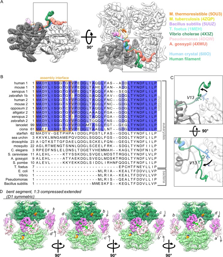
Figure 3—figure supplement 3. The vertebrate-specific N-terminus mediates IMPDH2 assembly of ATP-bound IMPDH2 filaments, in which individual protomers can extend or compress freely.

Figure 3—figure supplement 3.

(A) The conformation of the N-terminus seen in assembled filaments of human IMPDH2 is unique among solved IMPDH structures, including other human structures. Boxed regions correspond to views in panel C. (B) Sequence alignment of human IMPDH1 (human 1) and IMPDH2 (human 2) and other IMPDH homologues. (C) Comparison of N-terminus conformations from cryo-EM of assembled filament (green) and published crystallized IMPDH2 (blue, PDB ID 6I0O). (F) Rotated views of the cryo-EM density for the best resolved ATP/IMP/NAD+ bent structure, colored as in Figure 2. The asymmetric unit is a tetramer, and each of the four chains can be viewed by rotating incrementally by 90 degrees. Gray letters and arrows indicate chain symmetry mates and Bateman domain conformations.