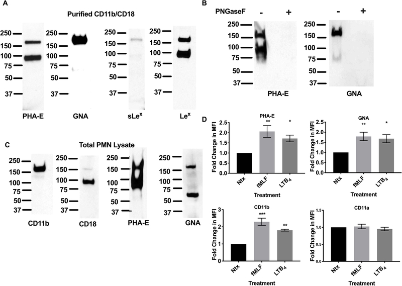
Figure 2. N-glycans recognized by PHA-E and GNA expressed on human CD11b.
(A) Purified CD11b/CD18 was immunoblotted using biotinylated PHA-E, GNA lectins or antibodies against sLex and Lex. (B) Immunoblots showing the CD11b/CD18 glycans recognized by PHA-E and GNA are N-glycan linked. (C) Total cell lysates from human PMN were immunoblotted with antibodies against CD11b and CD18 or biotinylated PHA-E and GNA lectins. (D) Changes in surface expression of CD11b/CD18, CD11a/CD18 and glycans recognized by PHA-E and GNA on human PMNs were analyzed by flow cytometry in non-stimulated (Ntx) PMNs and PMNs exposed to 100nM FMLF or 10nM LTB4 using FITC conjugated mAbs or lectins. Data are mean fluorescent intensities ± SEM (n=5). *, p<0.05, **, p<0.01, ***, p<0.001.

