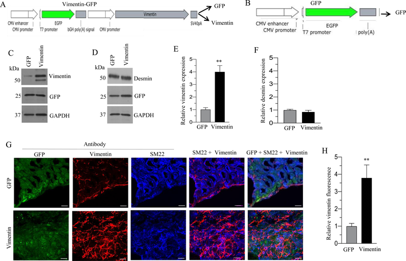
Figure 2. Adenovirus mediated overexpression of vimentin in murine BSM strips.
A: Schematic depiction of vimentin-GFP bicistronic adenoviral vector construct. B: Schematic depiction of GFP adenoviral vector construct. C-D: Murine BSM strips devoid of urothelium and submucosa were transduced with an adenovirus encoding GFP and vimentin for 48 h and the expression levels of vimentin, desmin and GFP proteins were determined by immunoblot analysis. GAPDH was used as a loading control. E-F: Quantification of immunoblot data. G: Sections prepared from murine BSM strips overexpressing vimentin and GFP proteins were stained with anti-vimentin, anti-SM22 and Alexa Fluor 488 labelled GFP antibody, followed by Cy3 and Cy5 conjugated secondary antibodies. Representative confocal images are shown. Scale bars = 10 μm. H: Quantification of confocal images data. Data are expressed as means ± SD (E, F & H), n = 5 mice in each group (E, F & H). **, P < 0.01 versus GFP expressing murine BSM strips.

