
Fig. 6.
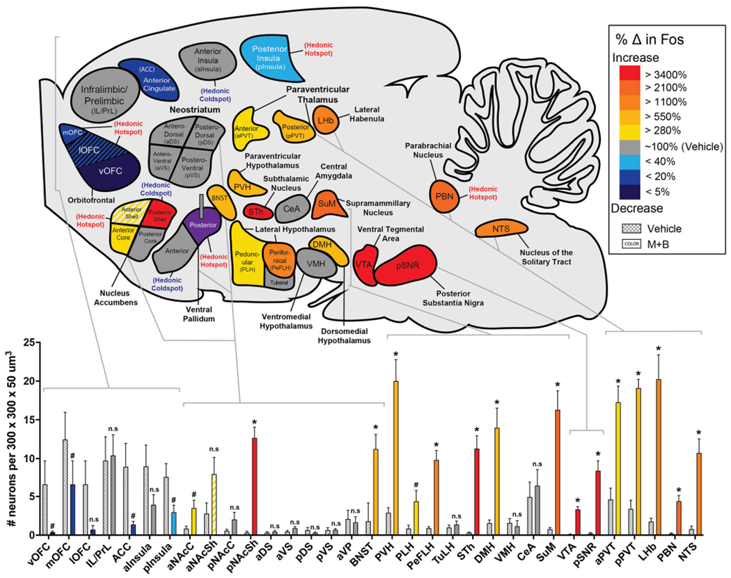
Whole-brain map of Fos activation changes recruited by excessive “disgust” during pVP inactivation. Sagittal map displays changes in Fos after muscimol + baclofen microinjections, compared with control baseline levels after vehicle microinjections. Colors reflect the significant percentage increase (red, orange, yellow) or decrease (blue, purple) in Fos levels recruited by excessive “disgust” after pVP inactivation compared with vehicle control baselines (100% = vehicle baseline, dark gray). Bars show absolute counts of Fos-expressing neurons in each corresponding brain structure. Increases in Fos expression were detected during “disgust” in the following structures: >280% in the anterior core of accumbens (aNAcC, p = 0.025), anterior shell of accumbens (aNAcSh, p = .087), anterior paraventricular nucleus of thalamus (aPVT, p < .001), and peduncular nucleus of lateral hypothalamus (PLH, p = 0.039); >550% in the basal nucleus of the stria terminalis (BNST, p =.002), paraventricular nucleus of hypothalamus (PVH, p < .001), and posterior paraventricular nucleus of thalamus (pPVT, p < .001); >1,100% in the perifornical nucleus of hypothalamus (PeFLH, p < .001), lateral habenula (LHb, p < .001), and nucleus of the solitary tract (NTS, p = .001); >2,100% in the supramammilary nucleus (SuM, p <.001) and parabrachial nucleus of the pons (PBN, p < .001); and >3,400% in the posterior shell of accumbens (pNAcSh, p < .001), subthalamic nucleus (STh, p < .001), ventral tegmental area (VTA, p < .001), and posterior substantia nigra (pSNR, p < .001). Decreases in Fos expression were detected during “disgust” in the following cortex regions: <40% baseline in the posterior insula cortex (pInsula, p = .035); <20% in the medial orbitofrontal cortex (mOFC, p =.024), and anterior cingulate cortex (ACC, p = .028); <20% in the lateral orbitofrontal cortex (lOFC, p = .104); and <5% in the ventral orbitofrontal cortex (vOFC, p = .026). Muscimol + baclofen microinjection did not cause significant changes in Fos expression in the infralimbic and prelimbic cortex regions of the anterior cingulate lobe (IL/PrL, p = .871), posterior core of accumbens (pNAcC, p = .156), anterior portion of ventral pallidum (aVP, p = .775), anterior insula cortex (aInsula, p = .126), any of the four quadrants of the neostriatum (aDS, p = .513; aVS, p = .513; pDS, p = .339; pVS, p = .934), tuberal nucleus of hypothalamus (TuLH, p = .495), central amygdala (CeA, p = .627), or ventromedial nucleus of hypothalamus (VMH, p = .659). #p < .05. *p < .005. (Color figure online)