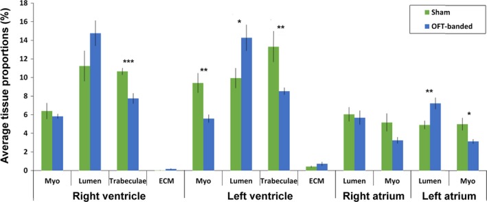
Figure 2.

Stereological analysis of tissue types contributing to different heart regions upon OFT‐banding. Reduction of trabeculae was found in both right and left ventricle of the OFT‐banded hearts, with an increase in left ventricular lumen. In addition, the myocardium was thinner in the left ventricle but normal in the right ventricle. Also, banding led to an increase of the lumen and a decrease of myocardium in the left atrium. ECM, extracellular matrix; Myo, myocardium. Sham, n = 6; banded, n = 7. Significant differences are indicated: *P < 0.05; **P < 0.01; ***P < 0.001. Error bars indicate SEM.
