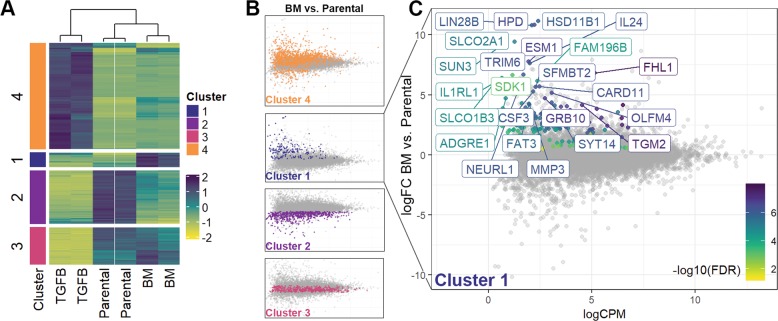
Fig. 1. Global characterization of gene expression following epithelial–mesenchymal plasticity.
a Dendrogram showing analysis of duplicate RNA sequencing analyses conducted on HME2 cells left untreated (Parental), treated with TGF-β1 for 4 weeks (TGFB) to induce a mesenchymal state, and TGF-β1-treated HME2 cells subcultured from a bone metastasis that formed subsequent to mammary fat pad engraftment (BM). As described in the “Materials and methods” section, gene expression changes were divided into four clusters based on differential expression between the three groups. b Cluster 1 was defined as genes whose expression did not change during TGF-β1-induced EMT but were significantly upregulated in the HME2-BM cells as compared to the HME2-parental cells. c Identification of an EMP signature of genes whose expression was significantly increased only after induction and metastatic reversion of EMT.

