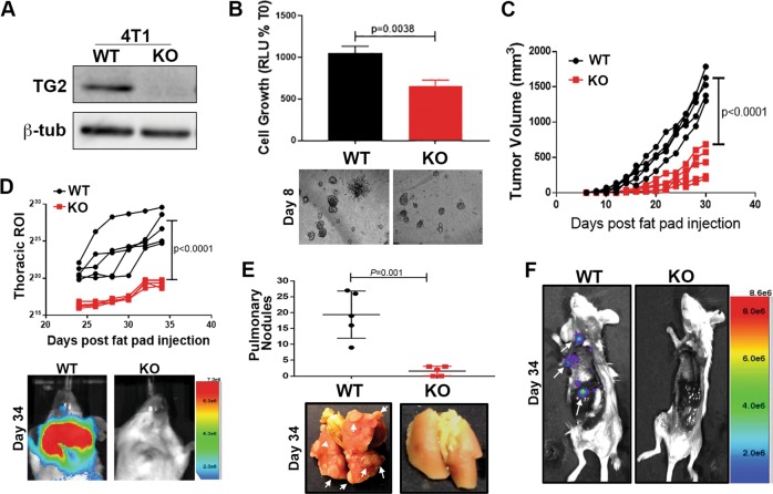
Fig. 4. Deletion of Transglutaminase-2 inhibits metastasis.
a Immunoblot for TG2 in control, wild-type (WT), and TGM2-deleted (KO) 4T1 cells. Expression of β-tubulin (β-tub) was used as a loading control. b Control (WT) and TGM2-deleted (KO) 4T1 cells were seeded under single-cell 3D culture conditions. Initiation of 3D outgrowth was quantified by bioluminescence. Data are normalized to the plated values and are the mean ± SD of three independent analyses resulting in the indicated p value. (below) Representative brightfield images of each 3D culture. c Control (WT) and TGM2-deleted (KO) 4T1 cells were engrafted onto the mammary fat pad and primary tumor growth was quantified by caliper measurements. Data are of individual mice taken at the indicated time points, resulting in the indicated p value. d Quantification of bioluminescent radiance from the pulmonary regions of interest (ROIs) at the indicated time points. (below) Representative thoracic bioluminescent images of control 4T1 (WT) TG2-deleted (KO) tumor-bearing mice. e Upon necropsy, the numbers of metastatic pulmonary nodules was quantified from control (WT) and TGM2-deleted (KO) 4T1 tumor-bearing mice. (below) Representative gross anatomical images of lungs from these groups. f Upon necropsy, the lungs and primary tumors of mice bearing control (WT) and TGM2-deleted (KO) 4T1 tumors were removed, and the carcasses were immediately imaged to visualize extra pulmonary metastases (arrows). A representative mouse from each group is shown. For d, e, data are the mean ± SE of five mice resulting in the indicated p values.

