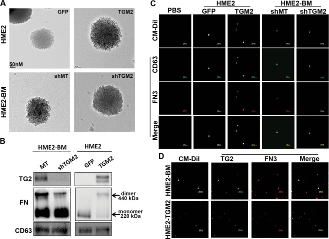
Fig. 5. Transglutaminase-2 promotes fibronectin crosslinking on the surface of extracellular vesicles.
a Transmission electron micrographs of extracellular vesicles derived from control (GFP) and TG2-overexpressing (TGM2) HME2 cells as well as control (shMT) and TG2-depleted (shTGM2) HME2-BM cells. b Immunoblot analysis of EVs derived from the HME2 and HME2-BM cells described in a. Differential expression of TG2 was verified in these EV lysates and correlated with covalent linkage of FN dimers that are insensitive to reducing conditions of the SDS-PAGE. CD63 served as a loading control. c Extracellular vesicle preparations derived from the cell types described in a were stained with CM-Dil (yellow) to verify the presence of lipid-containing particles. These preparations were also stained with antibodies specific for CD63 (green) and FN3 (red) and imaged using confocal microscope. The green (CD63) and red (FN3) channels were merged. A blank control sample (PBS) stained with the CM-Dil and appropriate secondary antibodies is also shown. d Extracellular vesicles derived from HME2-BM and HME2-TGM2 cells were stained with CM-Dil (yellow) and antibodies specific for TG2 (green) and fibrillar FN (FN3; red) and imaged using a confocal microscope. The green (TG2) and red (FN3) channels were merged. Scale bars on c, d are 500 nm.

