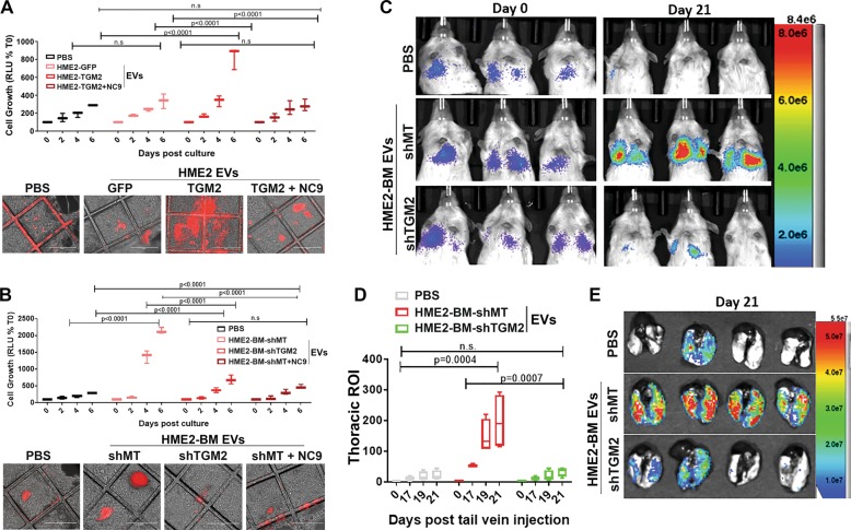
Fig. 8. Transglutaminase-2 enhances the ability of EVs to increase pulmonary colonization.
a, b Three-dimensional cultures of human pulmonary fibroblasts were treated with the indicated EVs as described in the “Materials and methods” section. MCF10-Ca1a cells stably expressing a firefly luciferase-dTomato fusion protein were subsequently added to these cultures, and their growth was longitudinally quantified by bioluminescence at the indicated time points. Data are mean relative luminescence units (RLU), normalized to the time zero (T0) reading, ±SE for three independent experiments completed in triplicate resulting in the indicated p values. Representative fluorescent and brightfield merged images showing Cala cell colonies (red) growing on the HPFs. c Mice were pretreated with the indicated EVs via intraperitoneal injections for 3 weeks prior to tail vein administration of the labeled MCF10-Ca1a cells described in a. Bioluminescent images of representative mice taken immediately (Day 0) and 21 days (Day 21) following tail vein injection of the MCF10-Ca1a cells. d The mean (±SE) bioluminescence values of thoracic regions of interest (ROIs) taken at the indicated time points. Data are normalized to the injected values, n = 4 mice in each group resulting in the indicated p value. e Mice were sacrificed at day 21, and upon necropsy, lungs were imaged ex vivo using bioluminescence to visualize pulmonary tumor formation.

