
Figure 7.
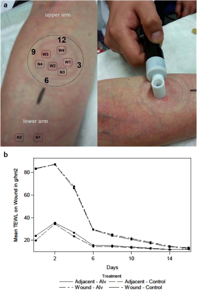
TEWL measurements of intact skin and superficial skin wounds. (a) With a suction blister device, a vacuum of 200 mm Hg was applied to the volar side of the lower arm to form blisters. TEWL measurements (right panel) were performed on non-dressed normal skin (N1 + N2), on normal skin covered by the occlusive wound dressing (N3 + N4) and on wounds. Wounds were measured clockwise starting from 3 o’clock (W1) up to 12 o’clock (W4). (b) The progression of mean TEWL of wound points. The wound points (W1–4) started with a high mean TEWL level compared to normal (N1 + 2) and adjacent skin points (N3 + 4). Wound levels increased from d0 to d2 and decreased very fast until d6 (dressing period) which indicates a fast healing process with both treatments. TEWL values were similar between treatments. n = 24, FAS cohort. Continuous line Alv, dotted line, control (Wilcoxon Signed Rank Test).