Figure 3.
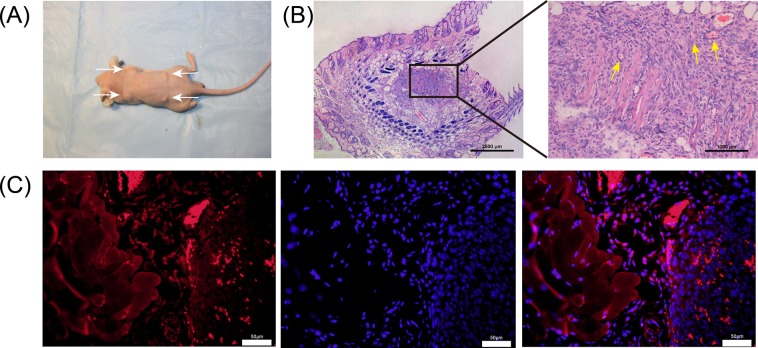
In vivo cell survival and differentiation of primary NSCs into neural-like cells. (A) NSCs/Matrigel complex was injected into nude mice (white arrows indicate the nodule location). (B) H&E-stained tumor sections from the transplanted NSCs showing good cell survival at 2 weeks post-transplantation (n = 5) (yellow arrows indicate new vessels). (C) Nodule sections were immunostained for β-tubulin III (red); nuclei were stained with DAPI (blue). Scale bars = ×200.

