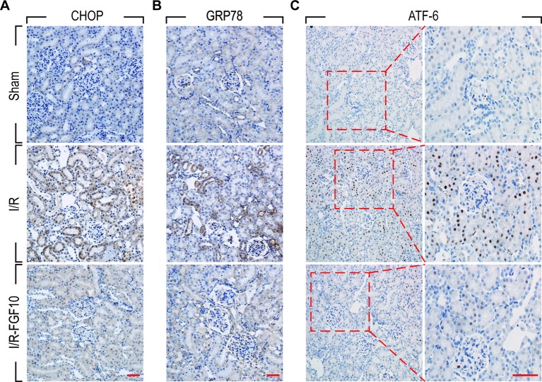
Figure 4.
Immunohistochemistry staining of ER stress relevant proteins in kidney tissues after reperfusion. (A) Immunohistochemistry staining for CHOP for renal tissues after 1 day of reperfusion. The expression of CHOP was significantly increased in the nucleus and cytoplasm of renal tubular epithelial cells after reperfusion, whereas FGF10 treatment reduced the expression of CHOP. (B) Immunohistochemistry staining for GRP78. FGF10 treatment reduced the expression of GRP78 in the cytoplasm of epithelial cells after reperfusion. (C) Immunohistochemistry staining for ATF-6. The expression of ATF-6 was significantly increased in the nucleus of renal tubular epithelial cells after reperfusion, whereas FGF10 treatment reduced the expression of ATF-6 compared to I/R alone. Panels are representative of five rats in each group. Scale bars represent 50 μm.

