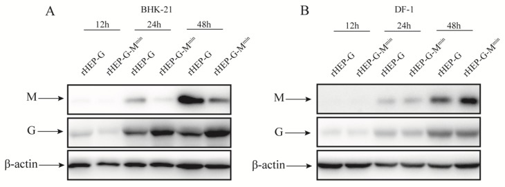
Figure 5.
Western blot analysis of M and G proteins. BHK-21 (A) and DF-1 (B) cells were infected with rHEP-dG, rHEP-dG-Mmin at an MOI of 3 and cell lysates were harvested at indicated time points post-infection. Western blot analysis was used to assess the relative expression levels of M, G, and β-actin.

