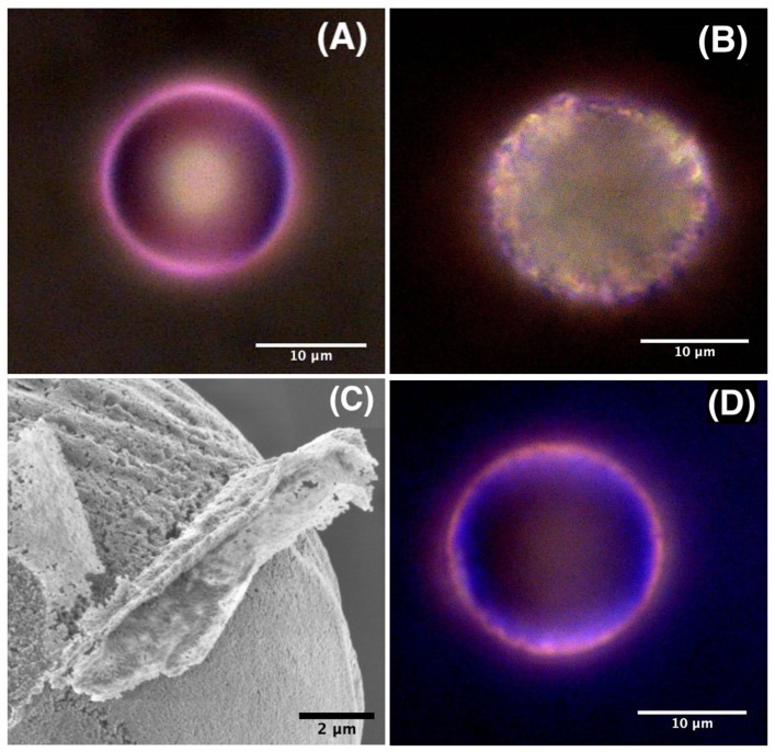
Figure 6.
Optical microscopy images of doped ZnO microspheres with RhB (A), Rh6G (B) and a RhB/Rh6G mixture (D); (C) High resolution scanning electron microscopy image of a ZnO microparticle showing the aggregation of ZnO nanocrystal building units of the microsphere and the resulting porosity of the material.

