
Figure 4.
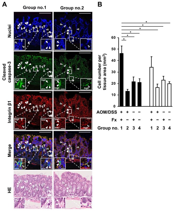
Detection of anoikis-like cells in colonic mucosal crypts in AOM/DSS mice with or without fucoxanthin (Fx) administration. The nuclei (blue fluorescence), cleaved caspase-3 (green fluorescence), and integrin β1 (red fluorescence) were observed by confocal microscopy. (A) Arrows show cells having negative/low expression (solid triangular arrows, anoikis-like cells) and high expression (open triangular arrows, non-anoikis-like cells) of integrin β1 plus high expression of cleaved caspase-3. HE, hematoxylin-eosin (HE)-stained sections. Long and short bars are 100 and 10 μm, respectively. (B) The number of anoikis-like cells (black box) and non-anoikis-like cells (white box) per tissue area (mm2) was determined by confocal microscopy. Mean ± SE (n = 5). Significant difference was performed by one-way ANOVA with a Tukey-Kramer post-hoc test. * p < 0.05.