Figure 3.
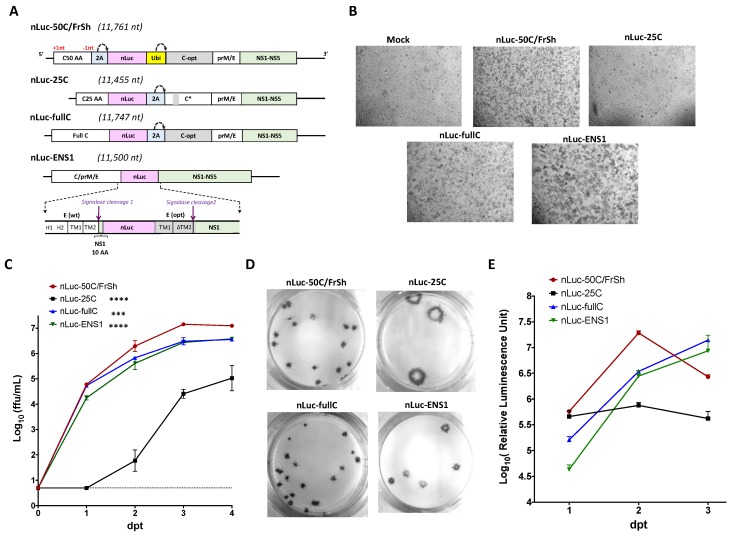
Construction and characterization of nLuc-carrying viruses in Vero cells. (A) Genetic organization of nLuc-containing viruses. ORF shifting and restoring mutations are highlighted as +1nt and −1nt, respectively. 2A: 2A protease sequence from FMDV; Ubi: ubiquitin sequence; gray boxes highlight codon-optimized sequences in the C and E genes of ZIKV. C* in nLuc-25C is a C opt gene with mutations in 14–17 AA codons. Dotted arrows represent the sites of cleavage by 2A protease or ubiquitin. (B) Microscopic evaluation (at 40× magnification) of CPE in Vero cells monolayer observed on day 6 after plasmid DNA transfection (dpt). (C) Growth kinetics of nLuc-carrying viruses in Vero cells after plasmid DNA transfection. Mean virus titer ± standard deviations in the samples that were collected daily from duplicate flasks was determined by titration on Vero cells. Dotted line represents limit of detection of the FFA (0.7 Log10(ffu/mL)). Differences between growth kinetics of nLuc-50C/FrSh and those of the other three constructs were compared using two-way ANOVA (**** p < 0.0001; *** p < 0.001). (D) Plaque morphology of nLuc-carrying viruses in Vero cell monolayer as revealed by immunostaining at 5 dpi. (E) Kinetics of luciferase activity following plasmid DNA transfection into Vero cells.

