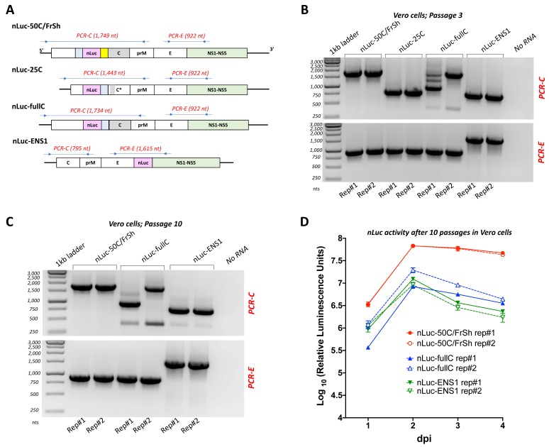
Figure 4.
Evaluation of the insert stability of recombinant nLuc-carrying viruses. To compare genome stability of ZIKV-carrying nLuc, blind passaging was performed for all four viruses in 12.5 cm2 flasks of Vero cells in duplicates, and RT-PCR reactions targeting the regions of nLuc insertion were carried out at multiple points during the experiment followed by sequencing. (A) Schematic representation of the positions of primers (represented by arrows) and expected lengths of corresponding RT-PCR fragments. (B,C) Agarose gel electrophoreses of RT-PCR fragments produced using viral RNA extracted from duplicate flasks of Vero cells (Rep#1 and Rep#2) after passage three (panel B) and ten (panel C). For each virus, two RT-PCR reactions were carried out (PCR-C and PCR-E, indicated in red), amplifying the region of nLuc gene insertion and selected region of ZIKV genome. Amplification of the viral regions that do not contain nLuc insertion serves as control of the expected band sizes after a complete deletion of the heterologous sequences in the nLuc-carrying viruses. Negative template control was included in each experiment and is shown in No RNA lane. Passaging of nLuc-25C was terminated after the third passage due to complete loss of nLuc insertion. (D) Kinetics of luciferase activity in Vero cells. Viruses recovered after the tenth passage in Vero cells were used for infection of Vero cells in 24-well plates at an MOI of 0.1. Measurements of nLuc activity in cell lysates were performed daily and data expressed as mean of standardized luciferase units ± standard deviations for the duplicate wells of infected Vero cells.

