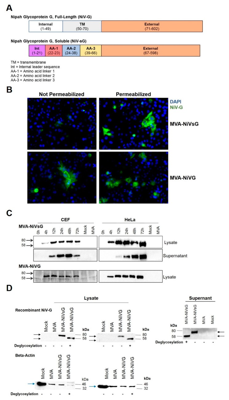
Figure 1.
Analysis of recombinant Nipah virus full-length glycoprotein G (NiV-G) protein produced by cells infected with MVA–NiVsG and MVA–NiV-G. (A) Schematic diagram of recombinant NiV full-length glycoprotein G (NiV-G) and NiV soluble glycoprotein G (NiVsG). Colored rectangles represent individual protein domains, and bracketed text displays the start and end of amino acid sequences of each domain. (B) Immunofluorescence staining of cells infected with MVA–NiVsG and MVA–NiV-G. HeLa cells were infected at MOI 0.05 with the above viruses for 16 h. MVA and mock infected HeLa cells were used as controls. Fixed permeabilized and fixed nonpermeabilized cells were immunostained with rabbit polyclonal antibody for NiV-G and the secondary antibody anti-rabbit Alexa Fluor 488. Nuclei were stained with DAPI solution. Panel shows representative pictures of fixed/permeabilized and fixed/nonpermeabilized infected HeLa cells at 40× magnification. (C) Western blot analysis of recombinant NiV-G proteins produced by chicken embryo fibroblasts (CEF) and HeLa cells infected with MVA–NiVsG and MVA–NiV-G. Lysates and culture supernatants were collected from cell cultures infected at MOI 5 with the above viruses, wild-type MVA or noninfected controls (mock). Samples were collected at indicated hours post-infection. Cell lysates and proteins were tested by immunoblotting using a NiV-G-specific polyclonal mouse antibody. Protein bands corresponding to the expected molecular weights of recombinant NiV-G and NiVsG protein (~65−70 kDa) are indicated. (D) Western Blot analysis of recombinant proteins produced by DF-1 cells infected with MVA–NiVsG and MVA–NiV-G at MOI 5 for 36 h. MVA and mock infected cells were used as controls. Cell lysates and culture supernatants were incubated with (+) or without (−) enzymes to deglycosylate proteins, analyzed by SDS-PAGE, and immunoblotted with a rabbit polyclonal antibody for NiV-G. Beta-actin was used as a loading control for lysates. Solid black arrow represents glycosylated recombinant NiV-G and NiVsG protein (~65–70 kDa), and dashed black arrow represents deglycosylated recombinant NiV-G and NiVsG protein (~58 kDa). Blue solid arrow represents beta-actin (~40 kDa). MVA: modified Vaccinia virus Ankara.

