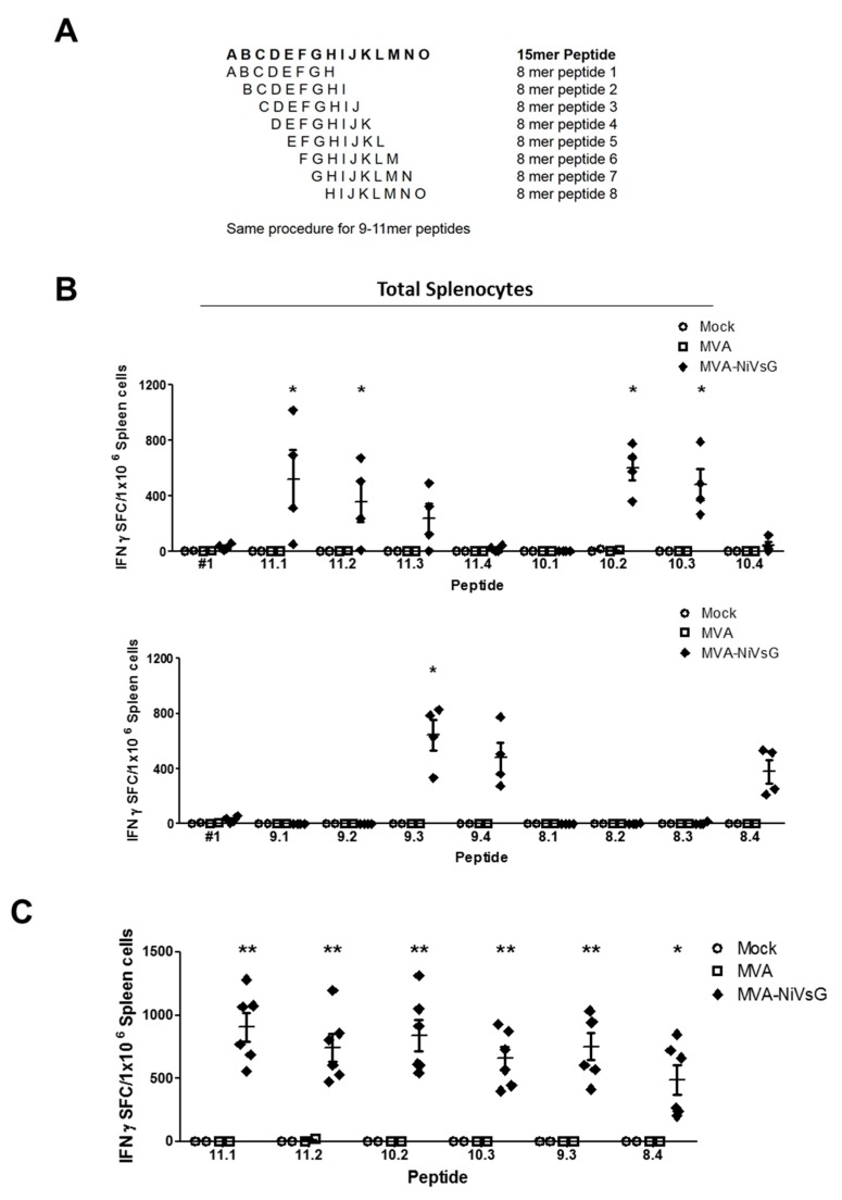
Figure 4.
Identification of H2-b-restricted T cell epitopes in the NiV-G protein. Groups of IFNAR−/− mice (n = 2–4) were immunized twice with MVA–NiVsG, MVA, or saline (mock) via the i.p. or intramuscular (i.m.) routes over a 21 day period. Spleens were collected and single cells suspensions were prepared 8 days after the final immunization. Total splenocytes were restimulated and measured by ELISPOT assay. (A) Schematic overview of 8–11mer overlapping peptide generation. The amino acid sequence of the positive 15mer peptide, #1, served to generate peptides with every possible 8–11mer sequence. Sequences were selected based on H2-b binding prediction results obtained from the SYFPEITHI database. (B) Mapping of H2-b-restricted 8–11mer overlapping peptides spanning the positive 15mer peptide #1 (YTRSTDNQAVIKDAL). Graphs show IFN-γ SFC of total splenocytes from i.p. immunized mice restimulated with peptide #1 and 8–11mer overlapping peptides. (C) Confirmation of positive H2-b-restricted peptides of NiV-G. Graph shows IFN-γ SFC of total splenocytes from i.m. immunized mice restimulated with the six most positive 8–11mer peptides (11.1, 11.2, 10.2, 10.3, 9.3, 8.4). Differences between groups were analyzed by one-way ANOVA. Asterisks represent statistically significant overall differences for a specific peptide. * p < 0.05, ** p < 0.01.

