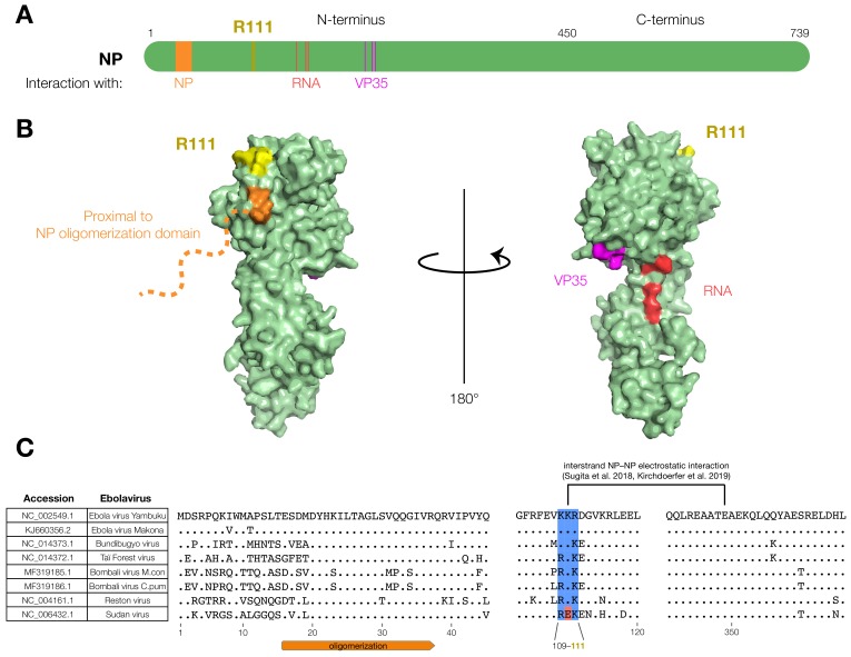
Figure 2.
The EBOV NP-R111 residue is unannotated, but could impact NP–NP interaction. (A) Schematic of NP. R111 (yellow) lies in an un-annotated region within the N-terminal lobe. Key residues of known NP interactions are highlighted; (B) Crystal structure (PDB #4YPI) of NP. Though the precise location of the oligomerization domain has yet to be determined by crystallography (orange dashed line), the R111 residue (yellow) is located on the same face as residues proximal to the oligomerization domain (orange: residues 39, 40), but opposite to the VP35 (magenta: residues 160, 171, 174) and RNA (red: residues 240, 248, 252) interaction interfaces; (C) Alignment of ebolavirus sequences. The basic residues at 109, 110, and 111 (blue), and a recently identified electrostatic interaction between K110 and E349 [28,29], are conserved in all known ebolaviruses except Sudan virus (SUDV, red).

