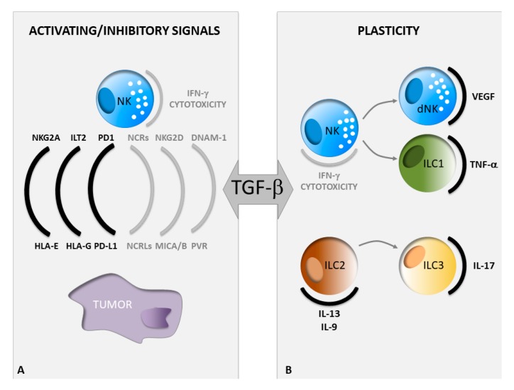
Figure 1.
Impact of TGF-β on ILC functions in cancer. (A) Phenotypic changes of both NK and tumor cells in a TGF-β rich tumor microenvironment. The grey or black color indicates a decrease or an increase in the expression levels of the depicted molecules, respectively. (B) TGF-β-driven conversion of one ILC subset into another, ILC, innate lymphoid cells; NK, natural killer.

