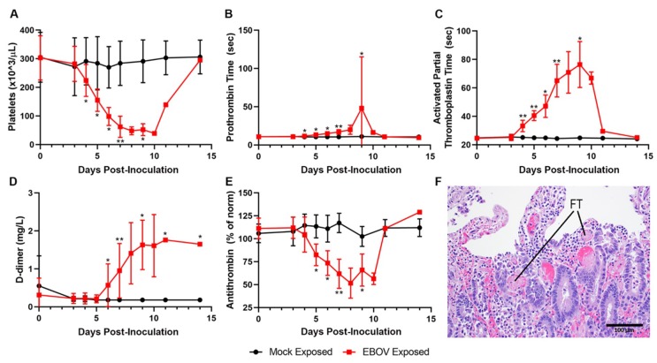
Figure 7.
Exposure to IM EBOV in rhesus macaques produces clinical pathology and histological alterations indicative of coagulopathy, including disseminated intravascular coagulopathy. Group means ± SD of: platelets (A); prothrombin time (B); activated partial thromboplastin time (C); d-dimer (D); and antithrombin (E). X-axes are truncated to highlight responses occurring from the first sampling point through the acute disease phase. * p < 0.05; ** p < 0.001. p-values are indicated for comparison of change-from-baseline values in mock- vs. EBOV-exposed animals on the indicated study day. (F) Small intestine, duodenal mucosa showing numerous intravascular fibrin thrombi (FT, two representative examples noted).

