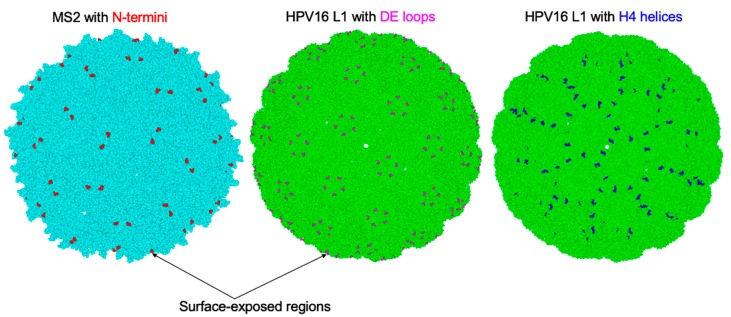
Figure 4.
Model cryo-electron microscopy images of icosahedral structures of MS2 (light blue) and HPV16 L1 (light green) derived from protein data bank (PDB) with PDB identification numbers 2WBH and 5KEP, respectively. The N-termini on MS2 coat proteins where L2 peptides have been inserted are shown in red (left image). The DE loops on HPV16 L1 coat proteins where L2 peptides have been inserted are shown in magenta (middle image). The H4 helices on HPV16 L1 coat proteins where L2 peptides have been inserted are shown in blue (right image).

