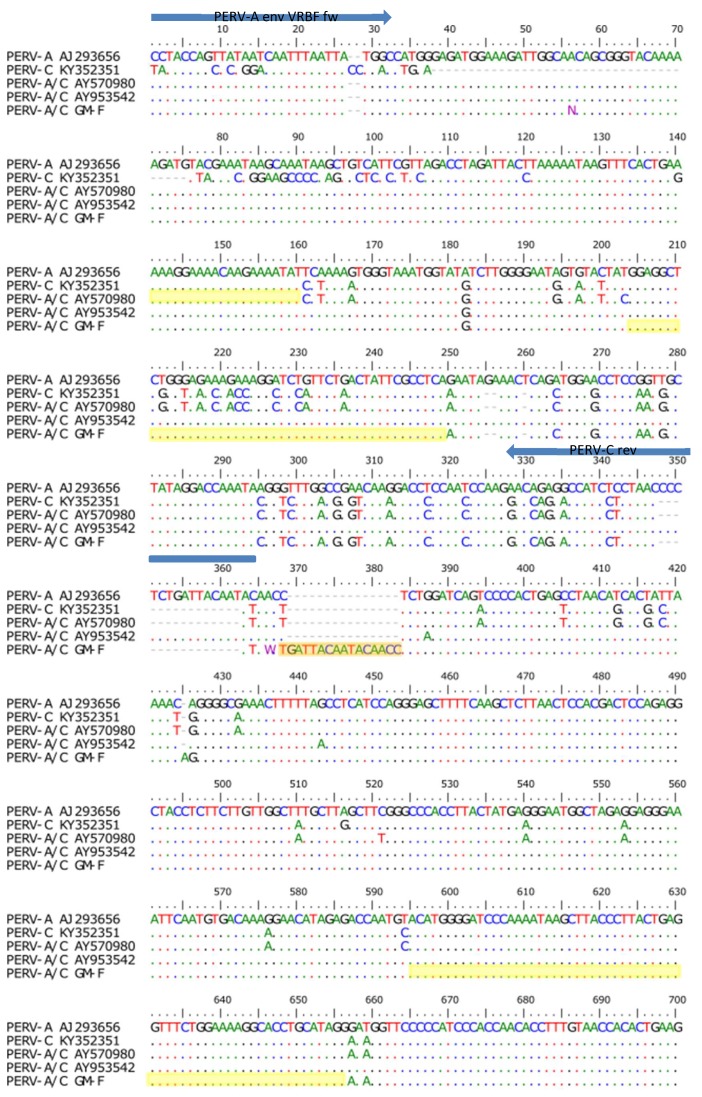
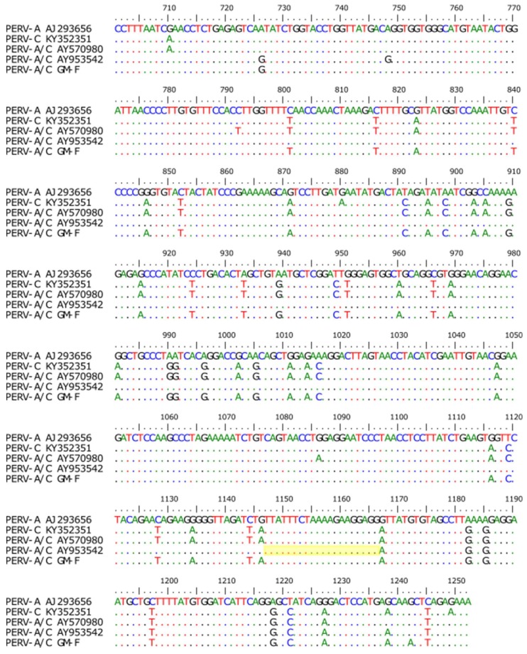
Figure 9.
Sequence alignment of PERV-A, PERV-C, and two PERV-A/C, the PERV-A 14/220 described by Bartosch et al. [37] (AY570980) and the PERV-A/C 50 analyzed in our laboratory (AY953542) [9,23,38], as well as the virus isolated from GöMP F (GM-F). The primer PERV-A VRBF (blue line) and the primer PERV-C-TMR (not shown, ranging from nt 1288–1263) (Table 2) were used for amplification and sequencing of the PERV-A/C env sequence. The primer PERV-C rev (blue line) was also used for the detection of PERV-A/C (Figure 4). The sequences marked yellow represent the potential regions of recombination, the orange bar indicate an insertion. N, any nucleotide; M, A or C, W, A or T.


