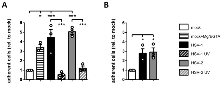
Figure 6.
HSV-2 induces fibronectin and ICAM-1 adhesion of mDCs. (A) Mock- (white bars), HSV-1- (MOI of 2, black filled bars), HSV-1 UV- (MOI of 2; 8 × 0.12 J/cm2, black striped bar), HSV-2- (MOI of 5; gray filled bars), or HSV-2 UV-infected (MOI of 5; 8 × 0.12 J/cm2, gray striped bar) mDCs were harvested 4 hpi. Mock controls were treated with or without Mg/EGTA. Cells were allowed to adhere on fibronectin-coated wells for 45 min. (B) Mature DCs were mock- (white column), HSV-1- (MOI of 2, black column), or HSV-2-infected (MOI of 5, gray column) and harvested 24 hpi. Cells were allowed to adhere to ICAM-1-Fc coated plates for 45 min. (A,B) Input conditions as well as adherent cells were quantified by measuring the β-glucuronidase activity. Changes in mDC adherence are shown relative to the mock condition (set to “1”).The experiment was performed three times with cells from different healthy donors, while each single condition was performed in quadruplicates. Error bars indicate ± SEM. Significant changes are marked by asterisks (*** = p < 0.001; *p < 0.05).

