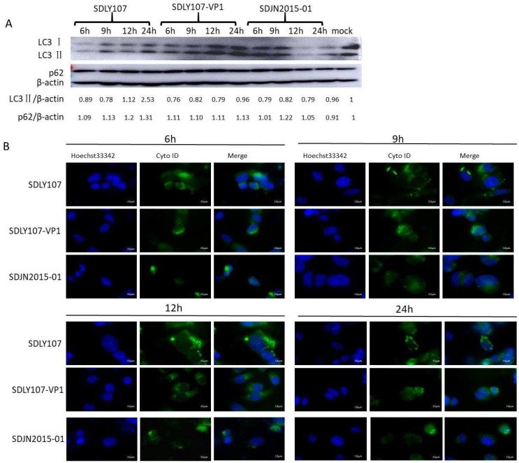
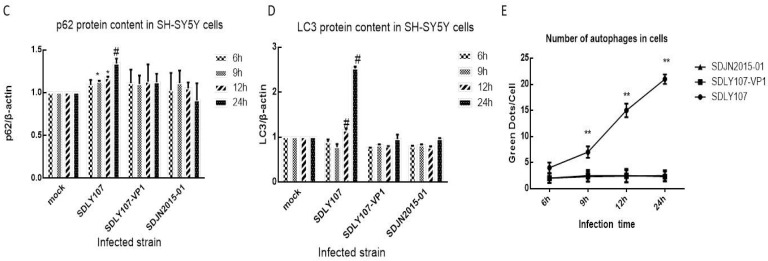
Figure 4.
(A) Western blot assay for the effect of recombinant virus SDLY107-VP1 strain, parental virus SDLY107 strain, and SDJN2015-01 strain on autophagy. SH-SY5Y cells were infected with EV71. Cell lysates were collected at 6 h, 9 h, 12 h, and 24 h. Uninfected cells served as the sham control. LC3 and p62 were examined by western blot. (B) The effects of the recombinant virus SDLY107-VP1 strain, parental virus SDLY107 strain, and SDJN2015-01 strain on autophagy were detected by immunofluorescent staining. Hoechst 33342 blue fluorescent dye stains the cell nucleus blue, and Cyto ID green fluorescent dye stains autophagous bodies green during autophagy. (C) The content of p62 protein in SH-SY5Y cells infected with the three viruses. (D) The content of LC3 protein in SH-SY5Y cells infected with the three viruses. (E) The number of autophages in SH-SY5Y cells infected with the three viruses. Autophagous bodies in each cell were counted, 100 cells per sample. The data represent means + SD from three experiments. * p < 0.05, compared with the mock group; # p < 0.001, compared with the mock group; ** p < 0.05, compared with the SDLY107-VP1-treated group.


