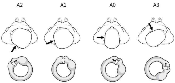
Figure 1.
Theoretical findings of the position of the cupula according to the head rotation angle during the head roll test in left persistent direction-changing positional nystagmus. The large and small arrows indicate the lesion side and direction of cupula movement, respectively. The left horizontal canal and cupula viewed from the front. A0 (supine position) cupula medial to lateral position. A1 (null plane) rotated 20–30° to the left (lesion side). Cupula parallel to the vertical axis. A2 (head turning to the left, lesion side) rotated an additional 40–50°. Cupula 40–50° downward from the vertical axis. A3 (head turning to the right, opposite side of the lesion) Cupula 90–100° to the vertical axis.

