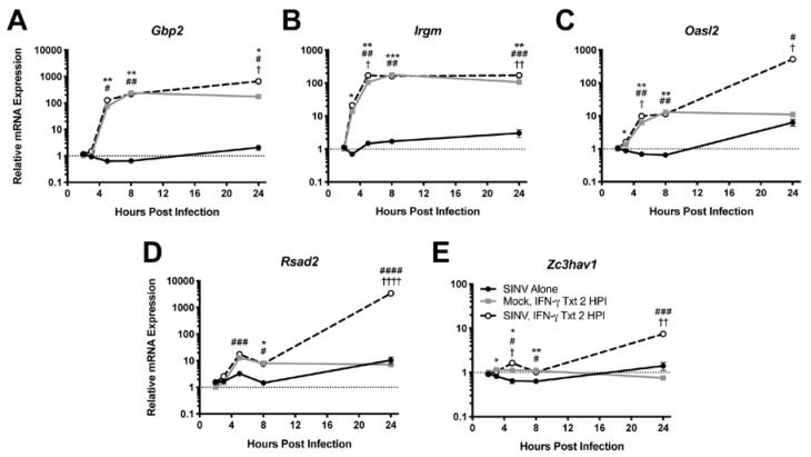
Figure 2.
Effect of IFN-γ treatment on ISG expression during SINV infection in vitro. ISGs selected for examination by qRT-PCR in dAP-7 cells infected with SINV at a MOI of 1 and left untreated (black circle and solid line), mock-infected and treated with 500 U/mL IFN-γ at 2 HPI (gray square and solid line), or SINV-infected at a MOI of 1 and treated with 500 U/mL IFN-γ at 2 HPI (white circle and black dashed line) included Gbp2 (A), Irgm (B), Oasl2 (C), Rsad2 (D), and Zc3hav1 (E) (n = 3 replicates per group; data are representative of two independent experiments and are presented as the mean ± SEM; dashed line indicates gene expression of untreated, mock-infected cells to which other groups were normalized; * p < 0.05, ** p < 0.01, *** p < 0.001, SINV Alone vs. Mock, IFN-γ Txt 2 HPI; # p < 0.05, ## p < 0.01, ### p < 0.001, #### p < 0.0001, SINV Alone vs. SINV, IFN-γ Txt 2 HPI; † p < 0.05, †† p < 0.01, †††† p < 0.0001, Mock, IFN-γ Txt 2 HPI vs. SINV, IFN-γ Txt 2 HPI, all by Tukey’s multiple comparisons test).

