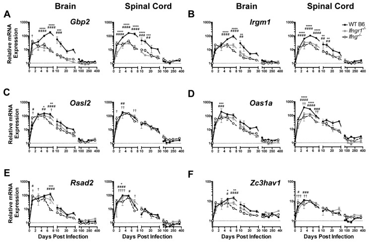
Figure 4.
ISG expression in SINV-infected mice with impaired IFN-γ signaling. Expression of Gbp2 (A), Irgm1 (B), Oasl2 (C), Oas1a (D), Rsad2 (E), and Zc3hav1 (F) were examined by qRT-PCR in the brains (left panels) and spinal cords (right panels) of WT (black circle and solid line), Ifngr1−/− (gray square and solid line), and Ifng−/− (white circle and black dashed line) mice (n = 3–6 mice per strain per time point; data are presented as the mean ± SEM; dashed line indicates gene expression of 0 DPI tissue for each strain to which other time points were normalized; * p < 0.05, ** p < 0.01, *** p < 0.001, **** p < 0.0001, WT vs. Ifngr1−/−; # p < 0.05, ## p < 0.01, ### p < 0.001, #### p < 0.0001 WT vs. Ifng−/−; † p < 0.05, †† p < 0.01, ††† p < 0.001, †††† p < 0.0001, and Ifngr1−/− vs. Ifng−/−, all by Tukey’s multiple comparisons test).

