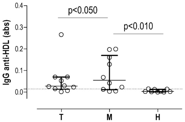
Figure 2.
IgG anti-HDL antibodies in tissue-conditioned media. IgG anti-HDL plasma levels (measured as absorbance) in tissue-conditioned media samples obtained from thrombus (T, n = 10) and media (M, n = 10) samples from AAA subjects and healthy (H, n = 10) aortas. Bars indicate medians, whereas whiskers represent 25th and 75th percentiles. Horizontal dotted line represents 90th percentile from healthy samples. Differences were assessed by Kruskal–Wallis tests (p = 0.0030) and p-values from Dunn–Bonferroni multiple comparisons tests are indicated in the figure.

