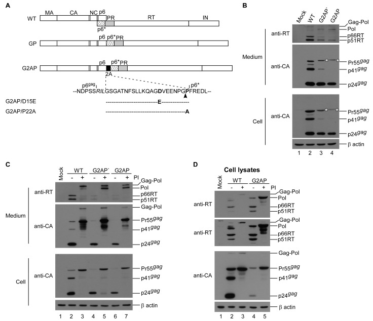
Figure 1.
Assembly and processing of HIV-1 Gag and Pol expressed from a single plasmid. (A) Schematic representations of HIV-1 Gag and Gag–Pol expression constructs. Indicated are the HIV-1 Gag protein domains MA (matrix), CA (capsid), NC (nucleocapsid), p6, 2A peptide sequence and pol-encoded p6*, PR, RT and IN. Arrowhead indicates 2A cleavage site. Altered or additional residues in italics. G2AP/D15E and G2AP/P22A are 2A substitution mutants, with a Glu substitution for Asp at position 15 and an Ala substitution for Pro at position 22, respectively. (B) G2AP assembly and processing. HEK 293T cells were transfected with designated construct. G2AP’ is identical to G2AP except for a Phe substitution for Ser-465 in Gag. Cells and supernatants were collected 48 h post-transfection and analyzed by Western immunoblotting. Membrane-bound proteins were initially probed with anti-RT serum prior to stripping and probing with an anti-p24CA monoclonal antibody. HIV-1 Gag–Pol, Pol, 66/51RT, Pr55gag, p41gag and p24gag positions are shown. Asterisks denote Gag-2A positions. (C) HIV-1 protease activity suppression increased G2AP virus production. HEK 293T cells were transfected with designated constructs. At 4 h post-transfection, equal amounts of cells were plated on two dishes and either left untreated or treated with saquinavir (an HIV-1 protease inhibitor) at a concentration of 5 μM. Supernatants and cells were collected 48 h post-transfection, prepared, and subjected to Western immunoblotting. (D) G2AP expressed Pol and Pr55gag at comparable levels. Cell lysates derived from panel C were probed with HIV-1 RT antiserum. For cellular background reduction, anti-RT serum was pre-incubated with membranes containing mock-transfected cell lysates. Middle panel: Images from longer exposures of the upper panel blot. Gag–Pol, Pol, 66/51RT and Gag protein positions are shown.

