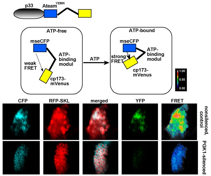
Figure 1.
Knockdown of cellular Pgk1 glycolytic enzyme inhibits ATP accumulation locally within tombusvirus viral replication compartments or replication organelles (VROs) in N. benthamiana. Top: Schematic representation of the FRET-based detection of ATP within VROs. The ATP biosensor, ATeamYEMK was fused to Tomato bushy stunt virus (TBSV) p33 replication protein. The dotted line indicates energy transfer between the modules. Bottom: Confocal microscopy images of VROs in plant cells show the low ATP level within the VRO when Pgk1 expression is silenced. Pgk1 mRNA level was knocked-down in N. benthamiana and the ATP level was detected via expression of the p33-ATeamYEMK biosensor. The YFP signal was generated via FRET. The more intense FRET signals are white and red (between 0.5 to 1.0 ratio), whereas the low FRET signals (0.1 and below) are light blue and dark blue. N. benthamiana plants were infected with TBSV, which replicates on peroxisomal membranes. CFP signal detects large TBSV VRO, which is also marked by the RFP-SKL peroxisomal marker. See further details in [33].

