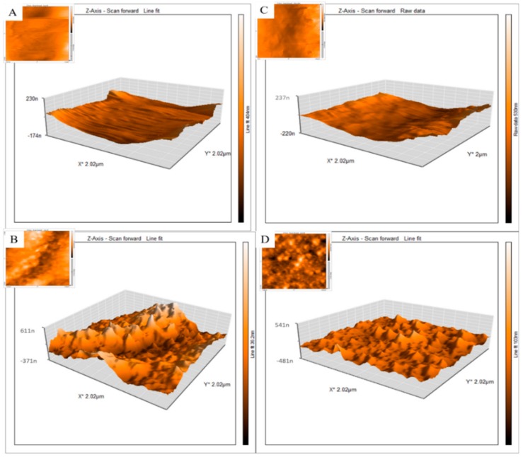
Figure 4.
2D and 3D atomic force microscopy (AFM) images of lab-on-a-chip fibrin scaffolds with and without AuNWs: (A) S1/Au−; (B) S1/Au+; (C) S2/Au− and (D) S2/Au+. As shown in Table 1 (S1 = stiffness 1, S2 = stiffness 2). Au+ = with AuNWs, Au− is without AuNWs as indicated in Table 1. n = 6 (at least) and p = 0.05. Significant differences were observed between (A,B) samples, as well as between (C,D).

