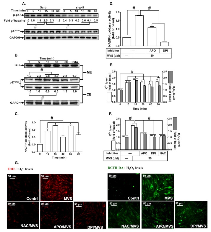
Figure 5.
Interaction between p47phox and Nox contributes to MVS-induced HO-1 expression. (A) HPAEpiCs were transfected with p47phox siRNA and then incubated with vehicle or MVS (30 μM) for the indicated time intervals. The levels of p47phox phosphorylation, total p47phox, and GAPDH were determined by Western blot using anti-phospho-p47phox, anti-p47phox, or anti-GAPDH antibody. (B) Cells were pretreated with MVS (30 μM) for the indicated time intervals. The cytosol and membrane fractions were prepared and analyzed by Western blot using anti-p47phox, anti-GAPDH, or anti-Gαs antibody. HPAEpiCs were treated with MVS for the indicated time intervals. The levels of (C,D) NADPH oxidase activity and (E–G) ROS generation (H2O2 or O2.–) were determined by ELISA or immunofluorescence (IF), reaching a maximal response within 15 min for Nox activity or 30 min for ROS. MVS stimulates Nox activity leading to ROS production, cells were pretreated with Nox or ROS inhibitors for 1 h, and then incubated with vehicle or MVS (30 μM) for (D) 15 min for Nox activity or (F,G) 30 min for ROS generation. MVS-induced Nox activity and ROS production were reduced by pretreatment with NAC (1 mM), APO (1 mM), and DPI (3 μM) in HPAEpiCs. Data are expressed as mean ± SEM from five independent experiments (n = 5). #P < 0.01 compared with the respective significantly different values as indicated.

