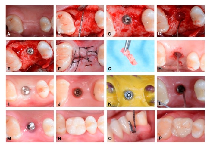
Figure 3.
Detail of a complete case performed in this study: from implant placement to the delivery of permanent restoration. (A–P). (A) Residual crest before incision; (B) bone explosion with periodontal probe to underline the residual bone; (C) implant just positioned; (D) gel inserted into the fixture (T0) ; (E) insertion of cover screw; (F) suture; (G) soft tissue biopsy; (H) gel inserted into the fixture after the removal of cover screw; (I) healing abutment placed; (J) soft tissue before the impression; during this stage the healing abutment was collected for the microbiological analysis (T2) ; (K) impression; (L) gel inserted before the positioning of abutment; (M) definitive abutment location; (N) provisional crown; (O) probing around implant; (P) definitive ceramic restoration delivery.

