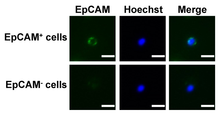
Figure 1.
Schematic representation for the fluorescence images of EpCAM+ and EpCAM− cells from individuals with advanced or metastatic HCC. The CD45+-depleted cell populations that were positive and negative for EpCAM were defined, respectively. Positive staining of Hoechst 33,342 (blue) indicates the presence of intact nucleated cells. Bar =10 µm.

