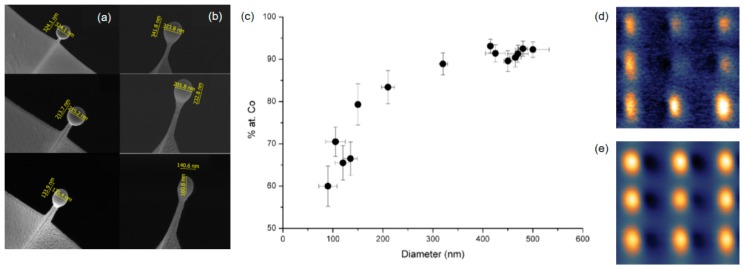
Figure 9.
FEBID-based ferromagnetic resonance force microscopy (FMRFM). (a) and (b) show differently sized Co nano-spheres (top down), fabricated on top of a high-resolution AFM cantilever in the top (a) and side views (b), revealing a close to spherical morphology. (c) shows the cobalt content of the beads as a function of the diameter, which has to be correlated with its magnetic properties. Additional measurements revealed a minimum diameter of 150 nm to exploit the full performance. (d) and (e) show FMRFM and MFM measurements, respectively, using a Co-AFM cantilever modified via FEBID. These measurements use an in-plane magnetic field, which aligns the sample magnetization. A microwave-frequency current is then introduced perpendicular to the magnetic field, but also in-plane. The latter decreases the quasi-static component of the magnetization, which impacts the magneto-static force between the sample and tip. Once the magnetic resonance is found, FMRFM reveals defects induced by chemistry or morphology, as provoked in this example by a different shape of the central element in the 3 × 3 array. This explains why that part is dark in FMRFM mode (d). In contrast, classical MFM measurements are unable to detect such defects, as shown in (e). Although powerful in its analytical capabilities, the lateral resolution is currently limited to about 90 nm, as that depends on the bead diameter. (a–c) were adapted and reprinted from Sangiao et al., Beilstein J. Nanotechnol. 2017 [117]. (d) and (e) were adapted and reprinted from Chia et al., Appl. Phys. Lett. 2012 [118].

