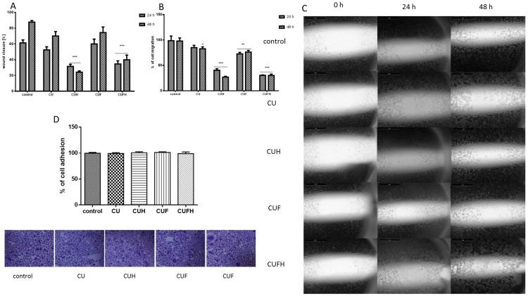
Figure 7.
The influence of 48 h of exposure of T. pratense extracts on wound healing (A) and migration (B) of HUVEC cells. Migration rates of HUVEC cells incubated with extracts at IC0 dosages into the free detection zones were photographed (×8) (C). Cells adhesion to the substrate was measured after staining with crystal violet; randomly chosen fields were photographed at ×200 (D). Control cells were not exposed to any compound but the vehicle; values are means ± standard deviations from at least three independent experiments; statistical significance was calculated versus control cells (untreated); * p ≤ 0.05, ** p ≤ 0.01, *** p ≤ 0.001.

