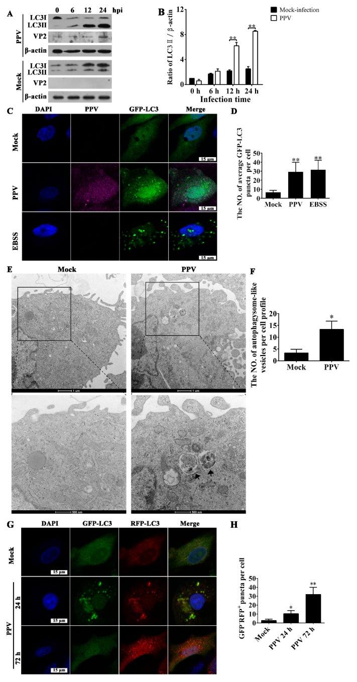
Figure 1.
Porcine parvovirus (PPV) infection triggers the accumulation of autophagosomes. (A,B) Porcine placental trophoblasts (PTCs) were mock infected or infected with PPV. The cellular and viral proteins indicated were evaluated via western blotting (A), and the ratio of LC3-II/β-actin in PPV-infected cells was calculated and analyzed (B). (C,D) PTCs were transfected with GFP-LC3 vector and then mock infected or infected with PPV for 24 h, followed by indirect immunofluorescence detection using antibodies against PPV capsid (ATCC CRL-1745), and corresponding Alexa fluo647-conjugated secondary antibodies. GFP-LC3 puncta formation was then observed under laser scanning confocal microscopy (C). DAPI (blue) was used to stain nuclear DNA; scale bar: 15 μm. The number of GFP-LC3 puncta in each cell was counted, with at least 50 cells were counted for each group. Next, the average number of GFP-LC3 puncta per cell was calculated (D). (E,F) Mock-infected and PPV-infected PTCs were processed and analyzed for the accumulation of autophagosomes via transmission electron microscopy (E). Black arrows indicate autophagic vesicles; scale bar: 1 μm and 500 nm, respectively. The number of autophagosome-like vesicles per cell profile was counted, and at least 15 cells were included for each group (F). (G,H) PTCs were infected with adenovirus RFP-GFP-LC3 for 12 h, and then were infected with PPV. The formation of autophagosomes and autolysosomes were observed (G). The number of GFP−RFP+-LC3 puncta in each cell was counted for at least 50 cells for each group, and the average number of GFP−RFP+-LC3 puncta per cell was calculated (H). The results are mean ± SD of three experiments. * p < 0.05 versus the mock-infected cells; ** p < 0.01 versus the mock-infected cells.

