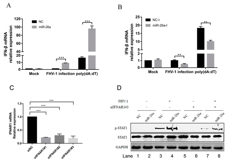
Figure 6.
miR-26a promotes the expression of type I interferon. (A,B) miR-26a enhanced FHV-1 or poly (dA:dT)-induced expression of IFN-β, while miR-26a inhibitors impaired IFN-β expression level induced by FHV-1 infection or poly (dA:dT) treatment. miR-26a mimics (A) or inhibitors (B) were transfected into F81 cells at a concentration of 80 nM or 160 nM for 24 h, respectively, then followed by FHV-1 infection at an MOI of 1 or transfection of poly (dA:dT) at a concentration of 1 μg/mL. Twenty-four hours after FHV-1 infection or poly (dA:dT) treatment, total RNAs were extracted for qRT-PCR to measure the mRNA expression level of IFN-β. The relative expression of IFN-β was normalized to that of cellular 18S RNA, and NC mimics and NC inhibitors served as negative controls. (C) The efficiency of IFNAR1 knockdown was evaluated by qRT-PCR. Three siRNAs targeting on feline IFNAR1 were transfected into F81 cells at a concentration of 60 nM, respectively. 36 h after transfection, cellular IFNAR1 mRNA expression level was tested by qRT-PCR. (D) F81 cells were co-transfected with siIFNAR1#3 (60 nM) and miR-26a mimics (80 nM) for 24 h followed by FHV-1 infection (MOI = 1) for another 24 h, and then the expression levels of p-STAT1 and STAT1 were identified by WB. All samples were independently repeated three times, and data are representative of three independent experiments. The significant differences are indicated as follows: NS > 0.05, ** p < 0.01, *** p < 0.001.

