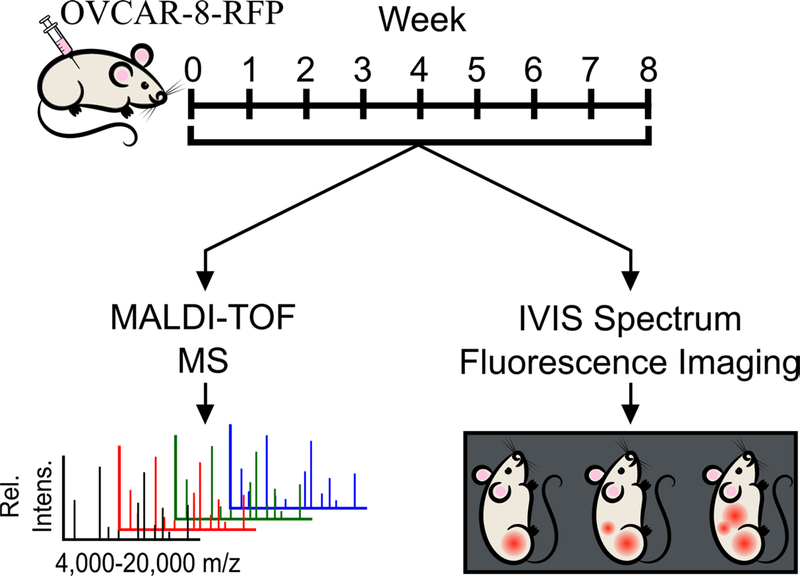
Figure 1.
Workflow of murine xenograft study. Female athymic mice were IP injected with OVCAR-8-RFP and tumors were allowed to develop over an eight-week period. Each week, mice were given vaginal lavages and were imaged using an IVIS imaging system to track tumor burden. Vaginal lavages were analyzed using MALDI-TOF MS to obtain mass spectra for statistical analyses.

