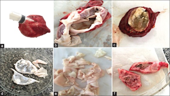
Figure-1.

Hydatid cyst of camel showing aspiration of the fluid from the cyst (a), opened evacuated hydatid cyst germinal layer of cyst wall that consisted of an outer thick fibrous layer and an inner thin germinal layer (b), opened hydatid cyst of cattle with semi-solid matrix (c), a germinal layer of hydatid cyst of camel liver (d and e), tissue from hydatid infected lung of sheep (f).
